Want help to write your Essay or Assignments? Click here
Mapping DNA using restriction Enzymes and electrophoresis
Mary Smith
Chem I – Sect C
10/12/20XX
Sediment in Water – Lab #3
Koeck (Edit this part with your details)
Abstract
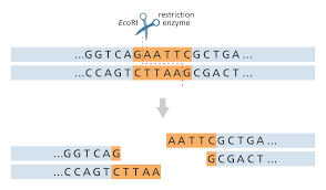
This laboratory report describes the experiment that was conducted using the restriction enzymes- restriction endonuclease- to manipulate the DNA molecules. The restriction enzyme has the capacity to recognize DNA sequences, and cleaving the DNA at that specific site. The endonuclease was used in conjunction with electrophoresis to map 2, 686 base pair of the pUC 19 plasmid. The plasmid cut into fragments, was separated using gel electrophoresis based on the charge and size.
Introduction
The first stage of DNA analysis for refined DNA such as gene expression and DNA sequencing is the construction of the DNA map. This has been done previously by scientists who used enzymes that are naturally occurring often referred to as restriction enzymes, to cut the large pieces of large molecule of DNA into small pieces. The fragments are then separated and sorted through the use of gel electrophoresis and the results obtained, they can be used to reconstruct the map of DNA molecule. This process is commonly referred to as mapping (Hughes and Moody, 2007).
This experiment was conducted using the restriction enzymes- restriction endonuclease- to manipulate the DNA molecules. The enzyme has the capacity to recognize specific DNA sequences, and cleaving the DNA at that specific site. The endonuclease will be used in conjunction with electrophoresis, which will be used to map 2, 686 base pair of the pUC 19 plasmid. The plasmid will be cut into fragments, which will be separated using gel electrophoresis based on the charge and size.
Want help to write your Essay or Assignments? Click here
Experiments (materials and methods)
Materials
Gel electrophoresis apparatus; Gel plates, comb to make wells, chamber cover, and chamber electrophoresis. Power supply with electrodes, deionized water, hot plate/ microwave, agarose, 250-Ml Erlenmeyer flask, and 100 ml graduated cylinder, pUC 19 plasmid DNA, restriction buffers, ice, restriction enzymes, molecular weight markers (lambda DNA), Ava II, Pvu II, gel loading dye (bromophenol blue), 15 1.5 Eppendorf’s, thermometer and metric rulers. Other common materials include, container that contains TBE solution, water bath (37 C), Floating rack, 60º C hot plate, cooler containing crushed ice, Polaroid camera with 667 Polaroid film, methylene blue stain, UV protective equipment, distilled water and non-frost free freezer.
Method
To make the Gel electrophoresis field, 1.0% of agarose was prepared as follows. To make 100 ml of gel, 1.0 g of the agarose was weighed and placed into the 250 ml glass beaker. 100 ML OF 1x TBE (Tris-Borate-EDTA) buffer was added. The mixture was heated in the hot pan for 30 seconds, shaking gently until all the agarose had melted completely. The solution was cooled and stored in a refrigerator.
The second day, pan was filled with water and was adjusted to 60 º C. The agarose was poured as follows; the agarose bottle that had been stored in day 1 was melted in the hot water bath. Firmly, the ends of the gel tray were sealed using a labelling tape, and a comb was placed on the slots, near the end of the tray, approximately, 40 ml of the agarose was poured in each of the tray, and was let cool for 15 minute to solidify.
During enzyme restriction stage, all enzymes and DNA aliquots were kept in ice. The four microtubes and reagents were labelled and stored as indicated below
| Reagents | Ava II | Pvu II | Control |
| 10 x Buffer | 4 µl | 4 µl | 4 µl |
| DNA | 4 µl | 4 µl | 4 µl |
| Pvu II | 0 µl | 2 µl | 0 µl |
| Ava II | 2 µl | 0 µl | 0 µl |
| Water | 30 µl | 30 µl | 30 µl |
The micropipette was set to collect 4µl and 4 10X restriction buffer to each of the tube. The similar process was followed to load 4.0 µl of DNA, but using a different tube, to the control tube, 32 µl of distilled water was added whereas in the other reaction tube, 30 µl of distilled water was added.
The microtubes caps were closed and were heated in the 55 ºC for 10 minutes and were placed immediately on ice for 2 minutes. 2 µl of the appropriate restriction enzymes were added as shown in the grid above. The microtubes caps were closed, tapped the tube gently to bring all the liquid to the bottom, and were incubated overnight at 37 C.
Want help to write your Essay or Assignments? Click here
To run the electrophoresis, the tubes were collected and placed in the ice tubes and the gel electrophoresis field was set up. The microtubes were heated in 60º C water bath for 3 minutes. 4 µl of the loading dye was added in each of the reaction tube. 20 µl of each sample was loaded in the well, and the current was turned on for 30 to 45 minutes. The gel was stained using the methylene blue solution in 0.1 TBE and was stained for 2 hours at room temperature. Observations were made and photograph was taken.
Results
There were several errors that we done during the preparation of the gel electrophoresis field. The first preparation did not gel, and the mistake could not be traced. This led to preparation of the second gel, which eventually worked perfectly. The loading of the DNA was also somewhat troublesome due to shaking of the hands, but I managed to pull it off, with the assistance of laboratory technician and my peers.
The following observations was made
1=2-log DNA (0.1-10 kb) molecular weight marker
2=pUC19 digested with AvaII, at 2464bp and 222bp
2 cuts =pUC19 digested with PvuII at 2364bp and 332 bp
4 cuts =pUC19 digested with AvaII and PvuII at 2464 bp, 2364bp, 332 bp and 222bp.
The control field had no fragments. Thebase pair at which the cuts occurred is almost the same number as those predicted by the pUC 19 maps.
Want help to write your Essay or Assignments? Click here
Discussion
The pCU19is a 26866 base pairs, and thus its kb can be estimated to be 2.7 kb. It is small, and a high copy number in E.coli plasmid, which contain pBR322 and M13MP19. It has multiple cloning sites, where each unique enzyme can cause restriction, thus facilitating the recombinant technology (Omoto and Lurquin, 2004).
The microtubes containing DNA were heated ath 60C to break hydrogen bonds at the end of the linear DNA. Addition of the dye also stopped the restriction reaction from taking place. After running the gel electrophoresis, DNA being negatively charged, it migrated towards the cathode, inform of bands of specific size. The controls had no fragments because the DNA was not digested by any restriction enzyme (Twyman, 2009).
The following observations were made; there were two cuts when pUC19 was digested with AvaII, at 2464bp and 222bp. This was also the same when pUC19 digested with PvuII, and the cuts were estimated to be at 2364bp and 332 bp. In double cleavage, a total of 4 cuts were observed when pUC19 was digested with AvaII and PvuII at 23640 bp, 2464 bp, 322 bp and 222 bp.
The control field had no fragments. The cuts are almost the same base pair number as those predicted by the pUC 19 maps. However, when compared the results from the attached pUC 19 map, the AvaII points should be digested at 1837 bp and 2059 bp, whereas, PvuII at 306 bp and 628 bp. The minor deviations could be associated with accuracy of recording the base pairs. The 4 cuts obtained by the double digests indicate that restriction enzymes recognize DNA sequences and cut them at that site (Twyman, 2009).
Want help to write your Essay or Assignments? Click here
Conclusion
The study objectives were achieved, restriction activity took place. This technique is a very important technique as it helps on interacts with the basics of cloning techniques and tools used in molecular biology.
References
Hughes, S. and Moody, A. (2007). PCR. Bloxham: Scion.
Omoto, C. and Lurquin, P. (2004). Genes and DNA. New York: Columbia University Press.
Twyman, R. (2009). Principles of Gene Manipulation and Genomics. John Wiley & Sons.
Want help to write your Essay or Assignments? Click here